Page 17 - SPEMD_58-2
P. 17
rev port estomatol med dent cir maxilofac . 2017;58(2):71-78 73
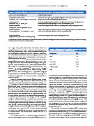
Table 1. Chemical composition of the orthodontic adhesives used in this study following manufacturers information.
Orthodontic adhesives (brand) Composition (% weight)
Compomer based adhesive 2 -Profenoic acid 2 Methyl, Phosphinicobis (oxy -2,1 -ethadilyl) Ester; Water; Mono HEMA
(Transbond Plus Color Change, 3M unitek, USA) phosphate; Tris [2 -(Methacryloyloxy)ethyl] phosphate
Resinous primer BISGMA (%5 -10); BISEMA (%10 -20); TEGDMA (%5 -10); Silane treated sílica (%2); Silane
(Transbond XT, 3M unitek, USA treated quartz (%70 -80); Diphenyliodonium Hexafuorophosphate
Resinous adhesive Uncured methacrylate ester monomers (%20 -38); Inert mineral fllers; fumed silica
(GrenglooT, Ormco Coporation, France)
Resinous adhesive Alkyl Dimethacrylate Resins (%60 -80); Ethyl alcohol (1 -5%); Barium Aluminoborosilicate
(Orthosolo primer, Kerr Corporation, USA) Glass; Fumed sílica (%2 -10); Sodium Hexafuorosilicate (%1 -5)
Resinous adhesive Uncured methacrylate ester monomers; inorganic fllers*
(Eagle No Drift, American Orthodontics, USA)
*Chemical composition is not available by the manufacturers information although elemental chemical analyses were performed in this study
to understand the shear bond strength results.
of 7 days. Then, teeth were stored in distilled water at a
temperature of 4 °C during 7 days for hydration prior to Table 2. Composition of Fusayamas artificial saliva used
as stock solution at pH 5.5 in this study.
bonding procedures. 9,10 Teeth were processed following the
technical specifcation ISO/TS 106 SC 11405:2003. After that, Compounds (g/l)
the teeth were cleaned with water and green stone at low
speed and then rinsed with water spray and air -dried under NaCl 0.4
oil -free airstream for 3 s. 9,10 The bonding surface was etched KCl 0.4
using 37% phosphoric acid (Octacid, Clarben S.A., pH < 2 at
2
2
20 °C) for 30s. Then, teeth were washed during 60 s and gen- CaCl .2H O 0.795
tly dried under airstream for 3 s. Teeth were divided corre- Na S.9H O 0.005
2
2
spondingly for bonding considering three orthodontic ad-
4
hesives: TP group: Transbond TM Plus Color Change/ NaH PO .2H O 0.69
2
2
Transbond TM XT Primer (3M unitek, USA); G group: Gren- Urea 1
gloo TM / Ortho Solo TM Primer, (Ormco Corporation, FRA); ED
group: Eagle No Drift TM (American Orthodontics, USA). The
chemical composition of the orthodontic adhesives is the interface. The remaining sixty were positioned with the
shown in Table 1. bracket base placed horizontally and then stabilized using a
Sixty -six stainless steel brackets (Master Series, Ameri- resin composite. For the shear tests, all teeth were embed-
can Orthodontics, USA) were bonded to enamel surface. The ded in PVC molds within self -curing acrylic resin to be at-
brackets bonding surface area was calculated as being 10.3 tached on a metallic holding device, that only the coronal
2
mm , using a 3D virtual model of the brackets by software region was exposed. Debonding was performed by axial
(SolidWorks, USA). One blinded and well -trained operator loading on the bracket wing, parallel to the bracket base,
performed the bonding procedure of the brackets to the using an universal testing machine (Instron 8874, 25kN; In-
enamel surfaces along the axis of the crown according to stron Corp., Norwood, Massachusetts, USA) at a crosshead
the manufacturer instructions. In all groups, excess com- speed of 0.5 mm/min (n = 10). 4,5,9 -16 Six samples (teeth with
posite material was removed with an explorer without dis- bracket) were randomly chosen for microscopic observation,
turbing bracket placement. Adhesives were light -cured by corresponding to one of each type of adhesive subjected or
continuous mode at 420 -490 nm using a LD -105 curing de- not to thermal clycing tests. After debonding, each sample
2
vice (1000 mW/cm , Monitex, China) for 30 s: on mesial and (n = 10) was inspected by optical microscopy (Axiotech, Carl
distal for 10 s each, over the bracker for 10 s. Afterwards, half Zeiss, USA) to evaluate the fracture pathways at magnifica-
of the samples were randomly chosen for thermal cycling tion ranging from x10 up tp x500. The residual adhesive on
tests (n =10). The thermal cycling tests were performed in the tooth was evaluated using the adhesive remnant index
Fusayamas artificial saliva solution (Table 2) at a tempera- (ARI) for 4 scores. 1,7
ture ranging from 5 up to 55 °C for 4000 cycles, according For scanning electron microscopy (SEM), samples were
12
to the ISO TR11450 (1994). Each cycle time corresponded to mounted in acrylic resin and cross -sectioned perpendicular to
45 s distributed in 15 s of dwell time in each bath and 15 s the bracket -adhesive interface plan. The cross -sectioned sam-
of transfer time. ples were wet ground on silicon carbide (SiC) papers down to
One sample from each six subgroup was randomly cho- 2500 mesh and polished using 1 m diamond slurry. The teeth-
sen for scanning electron microscopy (SEM) observation of -adhesive -bracket region of each sample was then inspected