
Page 26 - SPEMD_58-4
P. 26
214 rev port estomatol med dent cir maxilofac. 2017;58(4):212-218
xerostomia derived from suspected SS, according to the Euro- require plaque disclosing agents. Four surfaces per tooth were
pean criteria proposed by the American-European Consensus evaluated (vestibular, mesial, palatine and distal) and a score
10
Group. Patients were then asked if they had dental implan- of 0-3 was attributed to each of them, depending on the
ts. All patients were adults in full possession of their faculties amount of plaque visible: 0, no plaque; 1, plaque only regis-
and able to answer questions and participate in data registra- tered when passing a probe over the tooth surface; 2, moderate
tion. The study was conducted at the Dental Clinic of the Fa- accumulation of plaque deposits easily visible; 3, abundance
culty of Medicine and Dentistry, University of Murcia (Spain). of soft material and/or calculus. The final score was obtained
Patients were recruited consecutively over a period from Sep- by totaling the score on all the surfaces explored and dividing
tember 2014 to April 2017. Of these, 20 patients who had den- this figure by the total number of surfaces examined.
tal implants were invited to take part in the study. An SS diag- To evaluate the presence of gingival inflammation, the four
nosis was confirmed in seven cases according to the European dental surfaces (mesial, vestibular, distal and palatine) of all
criteria proposed by the American-European Consensus the teeth present in the oral cavity were evaluated using a
10
Group. The remaining thirteen patients presented xerosto- periodontal probe. The presence of bleeding on probing was
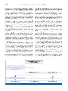
mia (Figure 1). marked by a plus sign and the absence by a minus sign. A
Patients with lymphoma, acquired immune deficiency percentage was calculated applying the formula: (number of
syndrome (AIDS), sarcoidosis and graft-versus-host disease, as surfaces presenting bleeding/number of surfaces with no blee-
well as patients in radiotherapy and/or chemotherapy, were ding) x 100. The value 0 was considered to indicate gingival
excluded. health.
A control group was created comprising patients with den- The probing depth was measured for each tooth present
tal implants who were willing to take part in the study but did in the oral cavity using a millimeter probe. Six points per too-
not suffer any symptoms of xerostomia or autoimmune disea- th were explored.The same six sites were also explored to find
se nor presented any salivary gland pathology. The study pro- the percentage of sites presenting epithelial insertion loss
tocol was designed to meet the criteria established by the De- greater than 3 mm. Gingival recession was obtained by mea-
claration of Helsinki for experiments involving human suring (in millimeters) the distance from the amelocemental
21
subjects and was approved by the University of Murcia’s Ethi- junction to the gingival margin. To calculate insertion loss,
cs Committee. the recession and pocket depth were added together for each
All patients were provided with full information about the site explored, and the index was obtained by applying the for-
purpose of the study and the procedures involved before giving mula: (number of sites explored with insertion loss > 3 mm /
their informed consent in writing to participate in the study. number of sites explored) x 100. Periodontitis was classified
The work followed the STROBE guidelines for case-control stu- according to Becks and Loe criteria, considering a value of
17
dies. A single clinician performed all oral and periodontal as- 0% as indicating no periodontal disease, 0-32% as slight perio-
sessments and evaluations of teeth and implants. dontitis, 33-66% as moderate periodontitis, and 67-100% as
Dental caries was assessed with the DMFT (decayed, missing, severe periodontitis.
filled teeth) index, according to the 1997 WHO parameters. 20 The following data were collected: the implant position
The Silness-Löe index was used to evaluate the bacterial (anterior when in the canine or incisor area and posterior
plaque on the gingival area around each of the teeth present when in the molar or premolar area), its antagonists (natural
in the oral cavity (except third molars); this technique does not teeth, prosthesis), its localization (upper arch, lower arch), the
INITIAL NUMBER OF PATIENTS
EVALUATED (n=89)
REASONS FOR EXCLUSION
Failure to meet inclusion criteria (n=65)
Refused to take part (n=2)
Other reasons ( n=1)
STUDY GROUPS (n =20) CONTROL GROUP (n=29)
Sjögren’s Syndrome (n=7) Xerostomia (n=13) CONTROL
Dental implants (n=29) Dental Implants (n=29) Dental Implants (n=140)
Figura 1. Patient flow diagram.